The human foot supports the body structurally, enables movement, supports the body’s weight, and withstands the strain of daily activities and athletic endeavors like running, jumping, lifting, and many other things. With 33 joints and more than 100 muscles, tendons, and ligaments, it is a complicated system. It’s incredible to think that each foot has 26 bones, or one-fourth of all the bones in the body.
The Item Nobody Wants
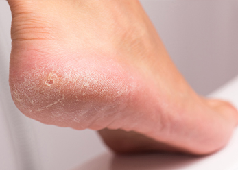
A small percentage of people (7–19%) are born with an additional bone on the inside of their foot, slightly above the arch. The auxiliary navicular, or os tibial externum, is a bone that develops from birth.
Since the auxiliary navicular bone is not a typical component of the foot, when the bone and posterior tibial tendon are inflamed, it can cause a condition that develops into a chronic syndrome.
The Beginning of Trouble
Between the ages of 8 and 15, while the bones are expanding and the cartilage is developing, the redness, irritation, and pain frequently manifest in patients. Sports and other activities become more demanding, which hurts the top of the foot where the protruding bone is located and damages the feet. Patients with flat feet may experience an aggravation of the problem. Adulthood can also see the pain’s manifestation. Causes of the inflammation:
- Trauma/foot or ankle sprain
- Irritation from shoes or other footwear rubbing against the extra bone
- Activity or overuse
Seek medical attention for your symptoms
If you or your teen experiences any of the following, schedule an appointment with American Foot for a checkup:
- A discernible bony protrusion directly above the arch on the inside of the midfoot
- Bony prominence edema and redness
- Generalized discomfort or throbbing in the arch and midfoot, typically felt during or after periods of activity.
Finding the syndrome’s cause
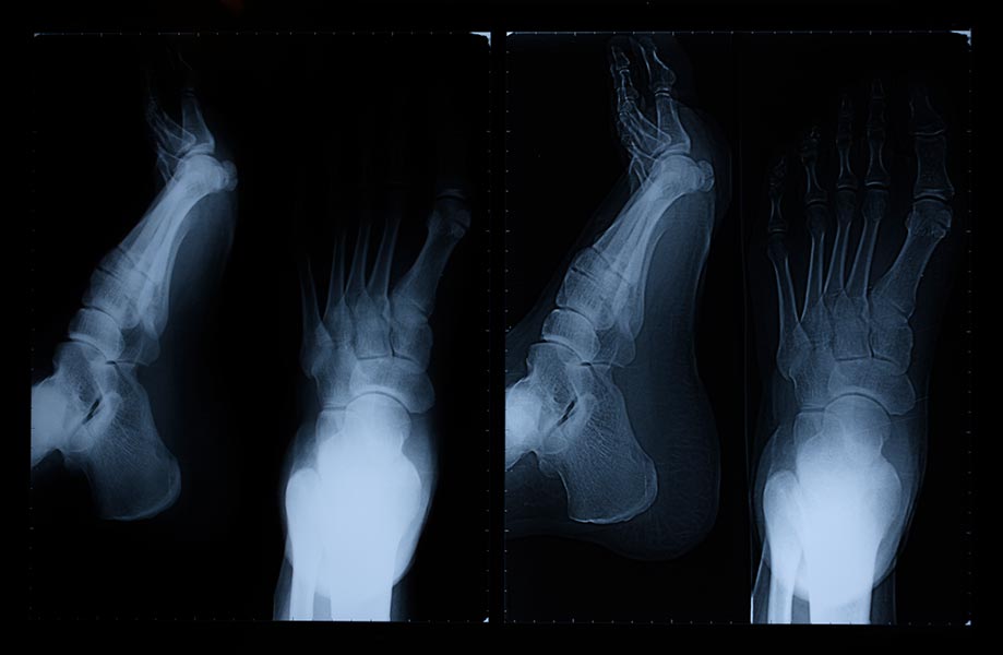
The Doctor at American Foot will examine the foot structure, joint and muscle strength, and range of motion in order to identify accessory navicular syndrome. To determine the intensity of the pain, he or she might press the bony prominence. In order to confirm the diagnosis, X-rays are used. For a more thorough analysis of the illness, an MRI or other sophisticated imaging studies may be used.
Non-surgical Methods of Therapy
Several non-surgical procedures can help alleviate the symptoms of auxiliary navicular bone condition, including:
- Immobilize—a cast or removable walking boot forces rest and enables the inflammation to subside. • Rest—avoid vigorous activity
Ice: It lessens edema. Avoid applying ice to the skin directly.
- Medication – NSAIDs (over-the-counter drugs), including ibuprofen, may be administered to treat pain and inflammation. Steroid therapy may be necessary in some circumstances.
- Physical therapy—builds muscle and aids in reducing inflammation. The right activities can be taught in therapy to stop symptoms from coming back.
- Orthotic devices: these offer arch support and decrease pressure that could result in irritation.
With careful attention, the foot discomfort may go away; nevertheless, because the bone is still present, it may start to hurt and feel tender once more. Surgery can be necessary if the pain comes again or lingers.
Surgical Approach
Your doctor at American Foot will operate on the auxiliary navicular bone following thorough consultation. Each patient’s surgical recovery period is unique. After surgery, the patient typically transitions from a cast to a cast boot walker after about 3 weeks. When the patient can walk in tennis shoes or after 4–6 weeks, physical rehabilitation starts. Usually, the patient can resume normal exercise after three months.